
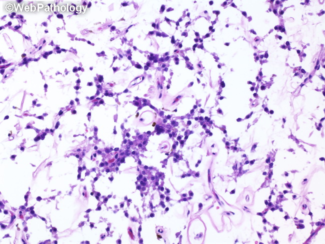
Leydig Cell Tumor with Partial Regression

slide 23 of 104


Comments:
The tumor cells are arranged in anastomosing cords in a background of delicate capillary network and fibroconnective tissue. Uninvolved testis is to the left. The patient was a 50 y/o male who was found to have a 0.7 cm intratesticular hypodense focus on ultrasound during workup for epididymitis. His serum tumor markers were within normal limits. The final diagnosis was - Leydig cell tumor with partial regression.
slide 23 of 104