
Alcoholic Cirrhosis

slide 22 of 34


Comments:
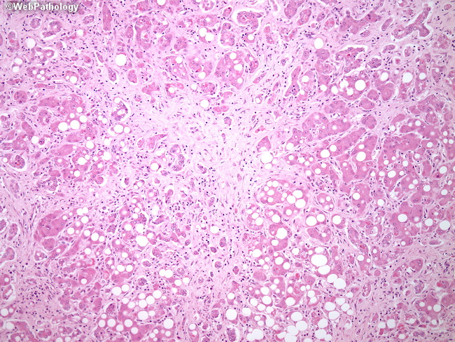
Fibrosis is an integral feature of alcoholic steatohepatitis and is typically seen in perivenular regions (acinar zone 3). Perisinusoidal stellate (Ito) cells deposit collagen as a result of Kupffer cell activation, release of platelet-activating factor, and neutrophilic infiltration. In severe steatohepatitis, the fibrosis extends to involve the remainder of the lobule as well as the portal tracts. This case of alcoholic cirrhosis shows marked steatosis with extensive hyaline sclerosis, which irreversibly distorts the parenchymal architecture.
slide 22 of 34