
Chordoma : Imaging


Comments:
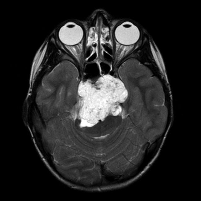
Chordoma - Imaging Studies: On imaging studies, chordomas appear as destructive, lytic lesions arising in the midline. They commonly extend into soft tissues, forming large well-defined masses, especially in sacrococcygeal region. The tumor is not mineralized but often shows bone fragments within and at the periphery. MRI is superior to other imaging modalities and shows lobular, septated, heterogenous mass with low intensity on T1-weighted images but bright lesions on T2 weighted images. There is heterogenous enhancement with contrast. Extension into soft tissues is better seen with MRI. The lesions appear radiolucent on CT. Sacrococcygeal tumors are difficult to identify on plain films due to obscuring effect of bowel gas shadows.Case History: The patient was an 8 y/o female with a mass at clivus. MRI showed a large retroclival prepontine mass infiltrating the posterior aspect of the clivus and compressing the brain stem posteriorly. It was hypointense in T1 and markedly hyperintense in T2 weighted axial image (shown here). The lesion was resected and confirmed to be a chordoma. Case courtesy of Professor Frank Gaillard, Radiopaedia.org. From the case rID: 7880 Note: Chordoma is quite uncommon in children. Patients under the age of 20 yrs. account for only about 5% of all cases. Tumors in children often tend to involve the base of the skull (as in this case) or upper cervical spine.