
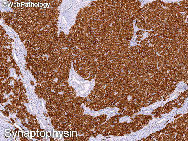
PanNETs : Synaptophysin


Comments:
Immunohistochemistry of Pancreatic Neuroendocrine Tumors (PanNETs): Both functioning and non-functioning PanNETs express neuroendocrine markers, including synaptophysin (integral membrane glycoprotein; strong diffuse staining), chromogranin (water-soluble acidic glycoproteins; focal and apical staining), NSE, CD56, and CD57. Even though the tumors may be non-functioning, they may still express peptide hormones or biogenic substances, including somatostatin, glucagon, and PP. About 40% of cases express more than one hormone. The specific hormone/s produced by the tumor are of no clinical or prognostic significance; therefore, immunohistochemical staining for specific peptide hormones is only of academic interest and is not required for diagnosis. Distinctive histologic patterns have been associated with specific hormones. Glucagon-positive NF-PanNETs show cystic change, reticular or trabecular pattern. Somatostatin-positive tumors have paraganglioma-like cell nests and/or glandular structures with psammoma bodies. Serotonin-positive cases have small cell nests and tubules in dense sclerotic stroma. Pancreatic neuroendocrine microadenomas (tumors <0.5 cm) are often positive for a single peptide hormone, usually glucagon followed by PP. Immunohistochemistry of PanNETs continues in the next image.