
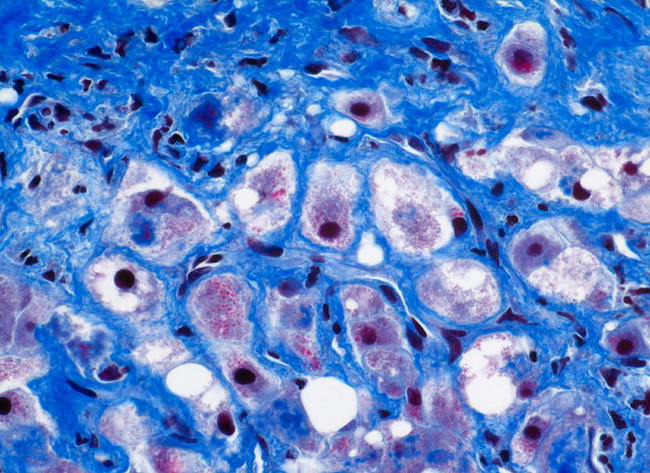
Alcoholic Steatohepatitis : Trichrome Stain


Comments:
Fibrosis is an integral feature of steatohepatitis and is typically seen in perivenular regions (acinar zone 3). Perisinusoidal stellate (Ito) cells deposit collagen as a result of Kupffer cell activation, release of platelet-activating factor, and neutrophilic infiltration. In severe steatohepatitis, the fibrosis extends to involve the remainder of the lobule as well as the portal tracts. Pericellular (�chicken-wire�) fibrosis is characteristic: strands of collagen surround damaged hepatocytes, forming a network that is highlighted by Trichrome staining (as seen here). Note the parenchymal inflammation and steatosis in this case. Pericellular fibrosis in the absence other features of steatohepatitis may result from a previous episode of steatohepatitis. Image Copyright: pathorama.ch.