
Extranodal Rosai-Dorfman Disease : Breast


Comments:
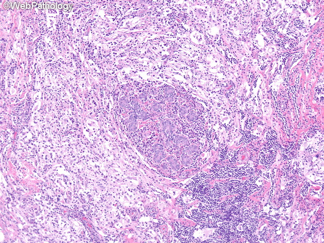
Microscopically, Rosai-Dorfman disease (RDD) of breast shows a dense inflammatory infiltrate of histiocytes (predominant cell), lymphocytes, and plasma cells in a fibrotic background. The infiltrate is not lobulocentric. Lymphoid follicles may be present. The histiocytes have large oval or round nucleus with open chromatin, single prominent nucleolus, and abundant eosinophilic or foamy cytoplasm. Some of the large histiocytes show emperipolesis, which refers to the presence of intact cells, usually lymphocytes, but sometimes plasma cells, red blood cells, and neutrophils within the histiocytic cytoplasm. Mitosis, necrosis, or granulomas are not seen. In contrast to nodal RDD, the extranodal sites show less conspicuous emperipolesis and more prominent fibrosis. RDD of breast has to be distinguished from diabetic mastopathy (which shows keloid-like fibrosis) and IgG4-related sclerosing disease (storiform fibrosis and obliterative phlebitis). RDD of breast has excellent overall prognosis. However, the disease may persist for months or years if not excised.