
Myxoid Leiomyosarcoma

slide 12 of 28


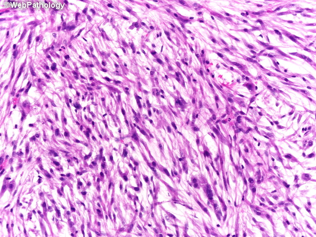
Comments:
The tumor consists of intersecting fascicles of spindle cells separated by abundant myxoid stroma. Other areas had more conventional morphology with plump spindle cells containing abundant eosinophilic cytoplasm, necrosis, nuclear anaplasia, and abnormal mitotic figures. The differential diagnosis includes inflammatory pseudotumor, post-operative spindle cell nodule, rhabdomyosarcoma, and sarcomatoid carcinoma.
slide 12 of 28