
Atypical Lipomatous Tumor/Well-Diff Liposarcoma : Imaging


Comments:
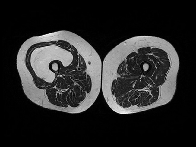
Atypical Lipomatous Tumor/Well-differentiated Liposarcoma - Imaging: Most ALT/WDL contain abundant mature fat and appear as fat density masses, but with less well-defined borders than lipomas. They may show mottled areas corresponding to fibrous or sclerotic component (see next image). This MRI (axial T2 weighted image) is from a 70 y/o female with a large thigh mass. In the vastus intermedius muscle of the right thigh, there is a large expansive mass that is isointense to subcutaneous adipose tissue in T1- and T2-weighted sequences. It measured 24 cm (craniocaudal) x 9 cm (transverse) x 7.5 cm (anteroposterior). The proximal and mid-distal portion of the tumor had some septa. The main neuro-vascular bundles did not appear to be compromised and there was no infiltration of the femur. The diagnosis of ALT/WDL was confirmed on histologic examination.Case courtesy of Valerio Giacalone, Radiopaedia.org. From the case rID: 97871