
Adenoid Cystic CA : Differential


Comments:
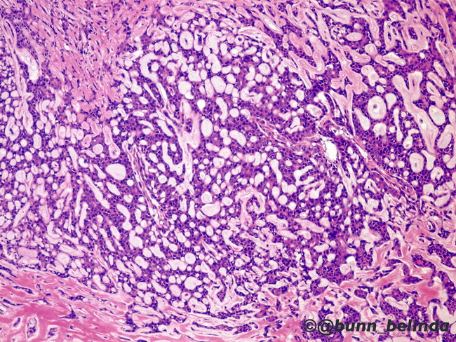
The differential diagnosis of adenoid cystic carcinoma (ACC) includes the following: Basal cell adenoma or Pleomorphic adenoma: Both entities can have overlapping histologic features with ACC. The presence of infiltrative margins, prominence of cribriform structures, and perineural invasion favor ACC. Features supporting pleomorphic adenoma include papillary architecture, true cyst formation, abundant chondromyxoid stroma, and squamous metaplasia. Polymorphous low-grade adenocarcinoma (PLGA): It almost never occurs in major salivary glands. PLGA has more abundant cytoplasm and uniform, round nuclei in contrast to ACC which has scant cytoplasm and hyperchromatic, angulated nuclei. Cribriform architecture and abundant hyaline material is more common in ACC. CD117 can be helpful in distinguishing between ACC (strongly positive) and PLGA (negative or weakly positive). In addition, diffuse S-100 positivity favor PLGA, whereas strong p63 positivity supports ACC. Epithelial-myoepithelial carcinoma (EMC): Features favoring EMC include clear cells, rare cribriform structures, rare basophilic myxoid material in microcystic spaces, and frequent branching glandular lumens. Based on the tumor location, basaloid squamous carcinoma and mucoepidermoid carcinoma can also enter in the differential diagnosis. The image shows nests and trabeculae of tumor cells punctured by round spaces filled with eosinophilic material and creating sieve-like appearance. Image courtesy of: Belinda Bunn, MD, University of Pretoria, Pretoria, South Africa.