
Adenomatoid Tumor

slide 10 of 41


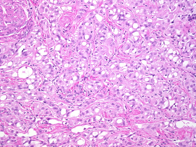
Comments:
The tumor is composed of low cuboidal or flattened cells arranged in solid cords, nests, glandular structures, cystically dilated spaces simulating vascular structures, or slit-like tubules. The stroma may have desmoplastic appearance and often contains smooth muscle, elastic fibers, and inflammatory cells. The cells have a small vesicular nucleus lacking any appreciable atypia, punctate nucleolus and eosinophilic cytoplasm. Intracytoplasmic vacuoles may be prominent and create a resemblance to signet ring-cells or fat cells. Differential diagnosis of adenomatoid tumor includes: metastatic carcinoma, malignant mesothelioma, and carcinoma of the rete testis.
slide 10 of 41