
Pleomorphic Adenoma

slide 57 of 110


Comments:
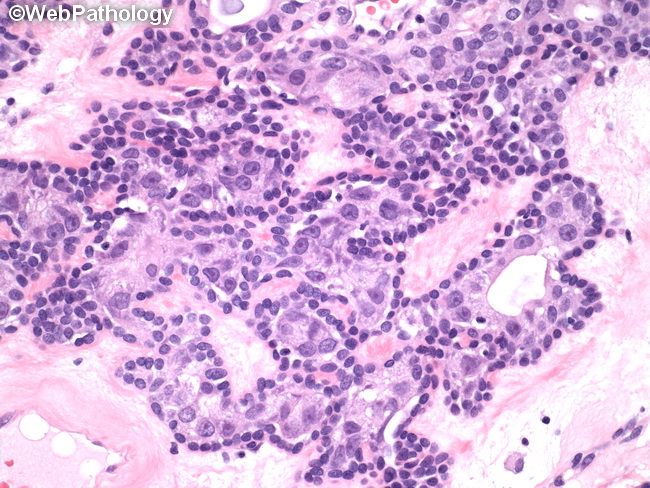
High magnification view of a pleomorphic adenoma demonstrates epithelial component arranged in a tubular structures lined by inner epithelial cell layer and outer myoepithelial layer in a background of eosinophilic myxoid matrix. The inner acinar or duct lining epithelial cells are flattened, cuboidal or columnar with moderate amount of cytoplasm and round nuclei. The outer myoepithelial layer is composed of spindle-shaped clear-appearing cells and may have plasmacytoid appearance due to eccentrically placed nuclei.
slide 57 of 110