
Myeloid Sarcoma : Skin


Comments:
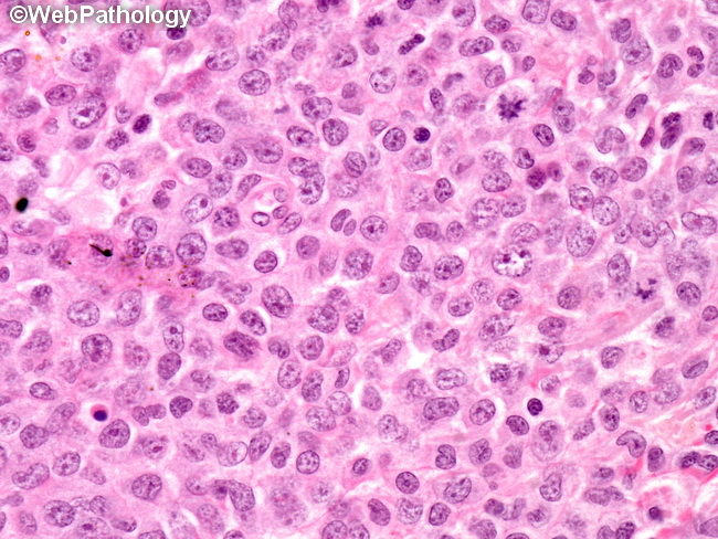
Cutaneous myeloid sarcoma: The cellular morphology depends upon the degree of maturation. Most cases show medium to large blasts with monoblastic or myelomonocytic appearance, arranged in solid sheets or infiltrating in single files (Indian file) in between collagen bundles. Eosinophilic myelocytes with their bright orange/red cytoplasm, when present, are easily recognized. Differential diagnosis of cutaneous myeloid sarcoma:
The main differential diagnoses include blastic plasmacytoid dendritic cell neoplasm, lymphoblastic leukemia/lymphoma, large B-cell lymphoma, and small round blue cell tumors of childhood. The distinction requires adequate clinical history and a broad panel of immunohistochemical markers. The greatest overlap in histologic features is with blastic plasmacytoid dendritic cell neoplasm which is positive for CD123 and BDCA-2 and negative for myeloid markers.