
Myeloid Sarcoma : Skin


Comments:
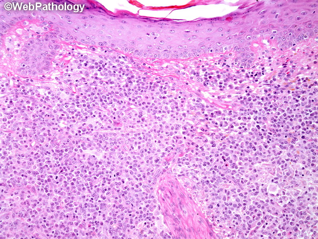
Cutaneous myeloid sarcoma is relatively uncommon, even though skin is one of the most frequently involved sites in acute myeloid leukemia. The clinical appearance of skin lesions is variable and includes erythematous or violaceous papules and nodules, firm infiltrative plaques, macules, and, less commonly, ulcers and blisters. Microscopically, there is nodular or diffuse leukemic infiltrate in the dermis and sometimes involving the subcutis. The epidermis is usually spared. Perivascular or periadnexal aggregation of tumor cells is common. The cellular morphology depends upon the degree of maturation. Most cases show medium to large blasts with monoblastic or myelomonocytic appearance, infiltrating, often in single files (Indian file), in between collagen bundles. Eosinophilic myelocytes with their bright orange/red cytoplasm, when present, are easily recognized. Skin lesions in myeloid sarcoma (leukemia cutis) are associated with 11q23.3 translocations and abnormalities of chromosome 16 such as inv(16).