
AITL : Morphology


Comments:
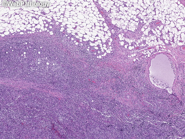
In the earliest stages of angioimmunoblastic T-cell lymphoma (AITL), the lymph node architecture is largely preserved (pattern 1) There are hyperplastic germinal centers with poorly-developed mantle zones. Any morphologic abnormalities are restricted to the interfollicular and pericapsular regions of the lymph node. The polymorphic cellular infiltrate usually extends beyond the node into the perinodal fat with sparing of the cortical sinuses (as shown here). The clues to the diagnosis of AITL include: increased vascularity (high endothelial venules), atypical T cells with clear cytoplasm, and proliferation of follicular dendritic cell network best visualized with CD21 immunostain. The atypical T cells are CD4+ and show aberrant expression of CD10, BCL, or PD1. They are located in the periphery of the germinal centers. The diagnosis of AITL in early stages can be made by confirming clonal rearrangement of T cell receptor genes.