
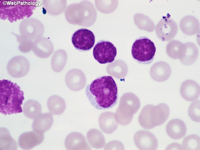
CLL in Peripheral Blood


Comments:
This image shows three small mature-appearing lymphocytes with condensed nuclear chromatin just above the center of the field. This is the typical appearance of lymphoid cells in peripheral blood in CLL. Two smudge cells are present along the left edge of the image. A prolymphocyte is seen just below the center of the field. It has slightly irregular nuclear contours, more dispersed chromatin, a prominent nucleolus and slightly more abundant cytoplasm. Proymphocytes usually make up less than 2% of the leukemic cell population in CLL. When they are more numerous (10% to 15% of neoplastic cells), the term atypical CLL is used. The atypical cases often show aberrant immunophenotype, specific cytogenetic abnormalities, cytopenias, refractoriness to therapy, more rapid disease progression, and overall worse outcome as compared to prototypic CLL cases. In some patients, an increase in the number of prolymphocytes may indicate impending clonal transformation. When the predominant cell type is prolymphocyte, a diagnosis of B-cell prolymphocytic leukemia should be considered.