
CLL/SLL in Spleen
Home
 Hematopathology
Mature B-cell Neoplasms - Part I
Chronic Lymphocytic Leukemia/SLL
CLL/SLL in Spleen
Hematopathology
Mature B-cell Neoplasms - Part I
Chronic Lymphocytic Leukemia/SLL
CLL/SLL in Spleen

slide 24 of 50


Comments:
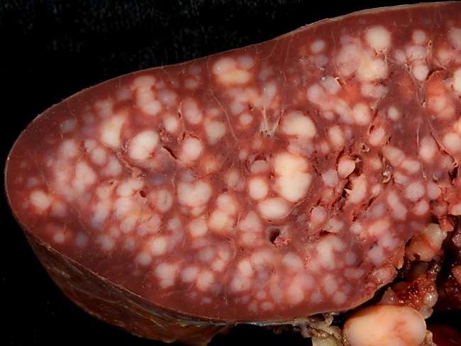
This 600-gram spleen is from an elderly lady who presented to the emergency room after an auto accident. There was no known history of CLL/SLL. At laparotomy, the spleen was found to be ruptured and was removed. The small whitish lesions scattered throughout the organ are nodular areas of white pulp expansion and represent splenic involvement in CLL/SLL. Meanwhile, the admission lab work showed a white blood count of 91,000/μL, almost all small round lymphocytes. Case courtesy of Ed Uthman, MD, Houston, Texas.
slide 24 of 50