
SLL in Lymph Node : Signet Ring Cell Pattern


Comments:
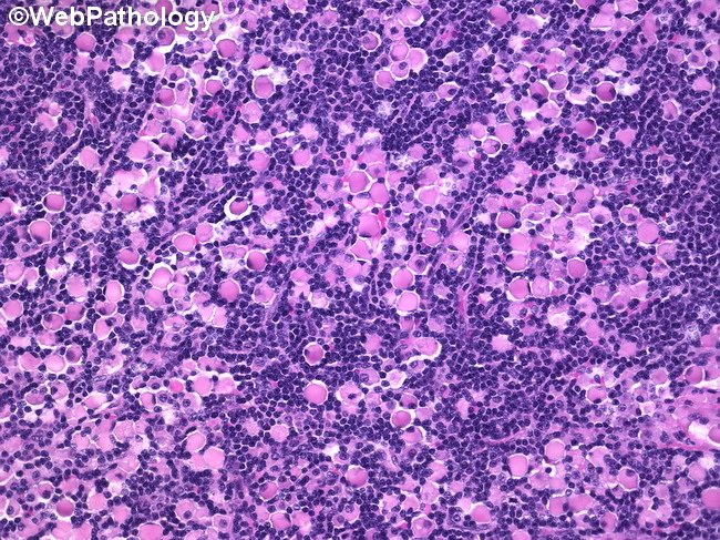
SLL with Signet Ring Cell Pattern: Sections of the lymph node show total effacement of nodal architecture by a diffuse proliferation of small uniform lymphoid cells with scant to moderate amount of cytoplasm, round or oval nuclei with coarse chromatin, and occasional punctate nucleoli. Admixed with the small lymphocytes are larger lymphoid cells with abundant cytoplasm containing rounded eosinophilic hyaline globules of different sizes. These globules displace and compress the nucleus to the periphery, imparting a signet-ring morphology. In many cells, the globules surround the centrally or eccentrically placed nucleus without distorting its shape. The signet ring lymphocytes formed large aggregates that had a predominantly sinusoidal distribution. Both the peripheral and the medullary sinuses were filled and distended by neoplastic signet-ring lymphocytes. The cytoplasmic hyaline globules were PAS-positive, diastase-resistant, and negative with mucicarmine stain. No mitoses were seen. Reference: Ramnani D, Lindberg G, Gokaslan T, & Albores-Saavedra J. Signet-ring cell variant of small lymphocytic lymphoma with a prominent sinusoidal pattern. Ann Diagn Pathol 3:220-226, 1999.