
Discoid Lupus - Oral Lesions
Home
 DermPath
Non-Neoplastic DermPath - I
Idiopathic Connective Tissue Diseases
Discoid Lupus - Oral Lesions
DermPath
Non-Neoplastic DermPath - I
Idiopathic Connective Tissue Diseases
Discoid Lupus - Oral Lesions

slide 25 of 59


Comments:
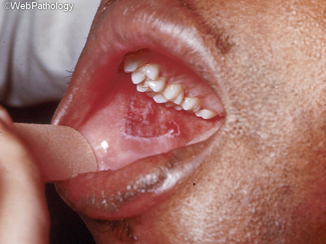
Oral lesions are seen in 20-25% of cases of discoid lupus erythematosus. The most commonly involved sites are vermilion border of the lower lip, alveolar process, and labial and buccal mucosae. The lesions are erythematous with white keratotic borders and may resemble atrophic lichen planus. Some lesions show erosions or ulcers. Chronic oral lesions are associated with an increased risk of squamous cell carcinoma.
slide 25 of 59