
Urachal Adenocarcinoma

slide 33 of 50


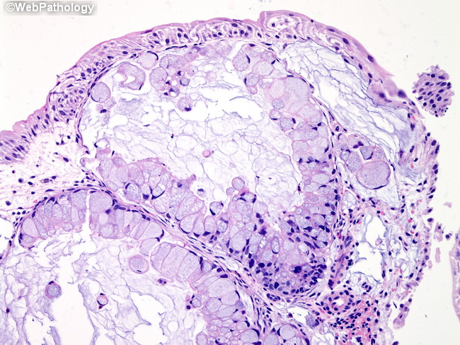
Comments:
This urachal adenocarcinoma presented in a young male with gross hematuria. CT-IVP showed a 3.0 cm mass with spotty calcification in the anterior-superior portion of the bladder near the dome. The biopsies shows mucinous adenocarcinoma in the lamina propria. Overlying urothelium was focally attenuated but uninvolved. In order for an adenocarcinoma to be labeled as urachal, following criteria must be met: location in the dome of the bladder; epicenter of the mass is in the muscularis propria (Detrusor muscle); absence of intestinal metaplasia or precursor lesions on the surface; no evidence of adenocarcinoma elsewhere (i.e. direct extension from a nearby structure or distant metastases must be ruled out).
slide 33 of 50