
Adenocarcinoma in Villous Adenoma
Home
 Genitourinary
Urinary Bladder
Glandular Lesions in Urinary Bladder
Adenocarcinoma in Villous Adenoma
Genitourinary
Urinary Bladder
Glandular Lesions in Urinary Bladder
Adenocarcinoma in Villous Adenoma

slide 17 of 50


Comments:
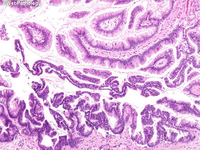
The upper half of this image shows typical morphology of a villous adenoma. The lesion in the lower half shows sufficient cytologic and architectural atypia to render the diagnosis of intramucosal carcinoma. One should be cautious in making the diagnosis of villous adenoma in a small specimen as any foci of coexisting adenocarcinoma may not have been sampled. It is also advisable to submit the specimen in its entirety to rule out malignancy.
slide 17 of 50