
Synovial Sarcoma : Radiology


Comments:
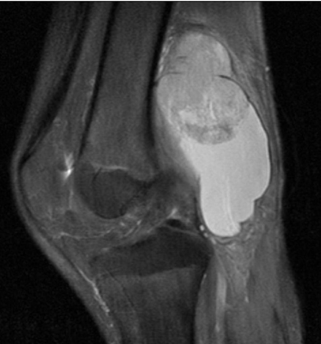
Radiologic Features of Synovial Sarcoma: Plain radiographs show round or oval, moderate density mass in the vicinity of a large joint. In a minority of cases, the nearby bone may show periosteal reaction, erosion or invasion. The presence of focal calcification or osseous metaplasia is seen as fine stippling or multiple small radiopacities in 15% to 20% of cases. Case History: Long-standing swelling in the popliteal fossa of a 13 y/o male. On ultrasound, the mass showed solid and cystic components. MRI with contrast (shown here) was performed depicting a mass with associated cystic component. The lesion is in the popliteal fossa immediately posterior to the femoral shaft. The diagnosis of synovial sarcoma was confirmed on surgery. Case courtesy of Dr Paresh K Desai , Radiopaedia.org. From the case rID: 10032