
Nodular Fasciitis : Breast


Comments:
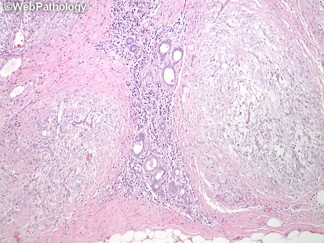
This is a case of nodular fasciitis involving the breast. The patient was a 25 y/o female who presented with a firm nodule in upper outer quadrant of her left breast discovered on self examination. An ultrasound showed a well-defined 2.0 cm hypoechoic mass. Fine needle aspirate revealed numerous spindle cells with mild cytologic atypia. The mass was excised. Grossly, there was a well-circumscribed, grey-tan myxoid lesion. Sections revealed a hypocellular lesion with a nodular configuration infiltrating in between ductal elements. It was composed of a uniform population of plump spindle cells with a tissue culture appearance (see next slide) in a myxoid stroma. In other areas, the cells were arranged in hyalinized stroma in short, intersecting fascicles with a vague storiform pattern. Extravasated red blood cells and scattered lymphocytes were present. Nodular fasciitis of breast typically arises in the subcutis, although it is within the breast parenchyma in this case (a rare occurrence). When it does occur deeply within the breast, it closely mimics breast malignancy clinically and radiologically.