
Mediastinal Germ Cell Tumor : Radiology
Home
 Mediastinum
Germ Cell Tumors of the Mediastinum
Mediastinal Germ Cell Tumor : Radiology
Mediastinum
Mediastinum
Germ Cell Tumors of the Mediastinum
Mediastinal Germ Cell Tumor : Radiology

slide 35 of 35

Comments:
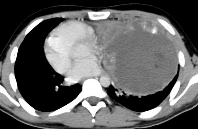
The patient was a 20 y/o male who presented with chest pain, dyspnea, and weight loss. A chest CT with contrast shows a heterogeneous mass filling most of the left thoracic cavum and displacing the mediastinum to the right. The tumor contained a single focal calcification and a few spots of fat densities. It invaded the pericardium, the thoracic wall and the diaphragm. CT-guided biopsy was performed. Histopathology report: Non-seminomatous mixed germ cell tumor composed of mature and immature teratoma, AFP-positive yolk sac tumour, and embryonal carcinoma. The patient received neoadjuvant chemotherapy before surgical resection. Case courtesy of Dr Roberto Schubert, Radiopaedia.org. From the case rID: 14131
slide 35 of 35