
Type B2 Thymoma : Differential


Comments:
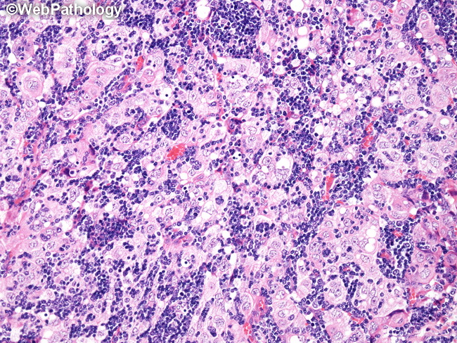
Type B2 Thymoma vs Type B1 Thymoma: Type B2 thymoma has greater epithelial cell density and epithelial cell clusters (as shown here) whereas the epithelial cells are fewer in number and scattered singly in type B1 thymoma. The presence of perivascular spaces is more typical of type B2 than type B1 thymoma; on the other hand, foci of medullary differentiation and Hassall corpuscles are more often seen in type B1 than type B2 thymomas. Type B2 Thymoma vs Type B3 Thymoma: Type B2 thymoma appears basophilic at low magnification due to its lymphocyte-rich background containing small clusters of epithelial cells. Type B3 thymoma is epithelial-predominant and lymphocyte-poor and therefore appears pink at low magnification. Type B2 thymoma vs Thymic Carcinomas: Type B2 thymomas may show focal anaplasia; however, they can be distinguished from thymic carcinomas by lobular architecture, perivascular spaces, TdT+ cells, and the absence of CD5/CD117 immunoreactivity. Type B2 thymoma vs T-Lymphoblastic Lymphoma (T-LBL): The blasts of T-LBL are monomorphic, atypical, and frequently infiltrate into mediastinal fat. Necrosis is frequently present. Epithelial stains such as CK19 and p63 fail to reveal epithelial cell network in T-LBL. In contrast, type B2 thymomas will show a population of CK19+ and p63+ epithelial cells and the T-cells are not monomorphic and lack atypia.