
Metastatic Tumors in Testis : Gross

slide 3 of 86


Comments:
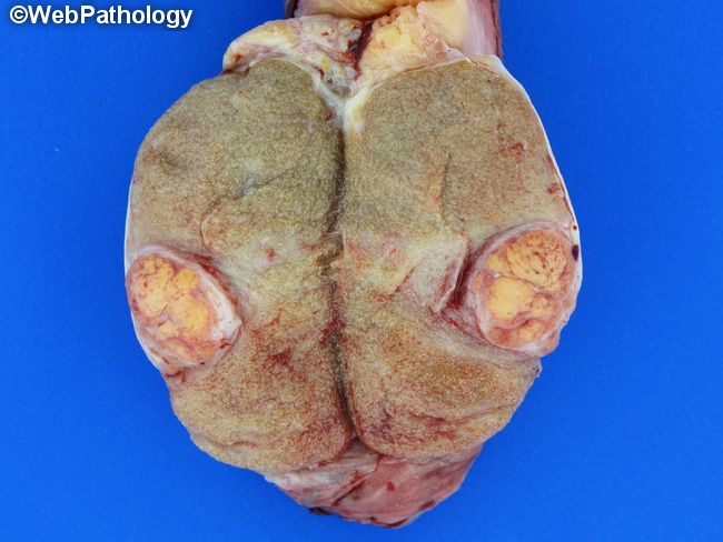
Gross Pathology: Testicular metastases can consist of solitary nodules (62% of cases), multiple nodules (17% of cases), or diffuse parenchymal involvement (21% of cases). The gross appearance of metastases depends upon the primary tumor type as shown here in a case of metastatic renal cell carcinoma in the testis. The well-circumscribed nodule has characteristic golden-yellow color that is typical of renal cell carcinoma, clear cell type (see close-up in the next image).
slide 3 of 86