
Choriocarcinoma : Differential Diagnosis


Comments:
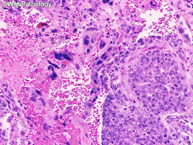
Differential Diagnosis of Choriocarcinoma (CC): Trophoblastic cells are present in many other germ cell tumors besides CC. However, they occur as single cells or small aggregates of syncytiotrophoblasts. The biphasic pattern of CC (cytotrophoblasts capped by syncytiotrophoblasts) is not seen. Embryonal carcinoma (EC) can have large atypical cells with smudged hyperchromatic nuclei resembling syncytiotrophoblast. Features favoring EC include: lack of hemorrhage, absence of hCG immunoreactivity, and positivity for OCT3/4. Monophasic choriocarcinomas should be distinguished from seminomas and yolk sac tumor with solid pattern. Features supporting choriocarcinoma include: greater degree of pleomorphism, diffuse immunoreactivity for hCG and negativity for AFP and OCT3/4. Placental site trophoblastic tumors show diffuse immunoreactivity for human placental lactogen and lack the biphasic appearance of choriocarcinoma. This image from a testicular choriocarcinoma shows a sheet of intermediate trophoblasts on the right and several syncytiotrophoblasts with smudgy hyperchromatic nuclei on the left.