
Chondroblastoma

slide 2 of 15


Comments:
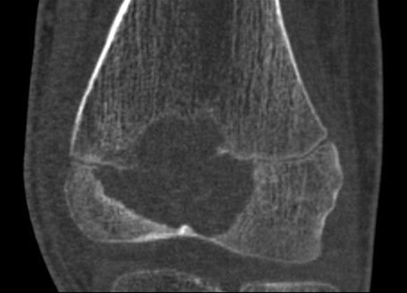
CT confirms the plain film appearance of the previous image, revealing a sharply demarcated epiphyseal lucent lesion but with faintly sclerotic margins. It transgresses the growth plate into the anterior part of the metaphysis. There is no periosteal reaction. The presence of a lucent lesion involving both sides of an open epiphyseal plate (as seen here) is diagnostic of chondroblastoma. Case produced with permission, courtesy of Dr. Frank Gaillard. Radiopaedia. Complete case is here.
slide 2 of 15