
Malignant Fibrous Histiocytoma of Breast
Home
 Breast
Rare Breast Tumors
Malignant Fibrous Histiocytoma
Malignant Fibrous Histiocytoma of Breast
Breast
Rare Breast Tumors
Malignant Fibrous Histiocytoma
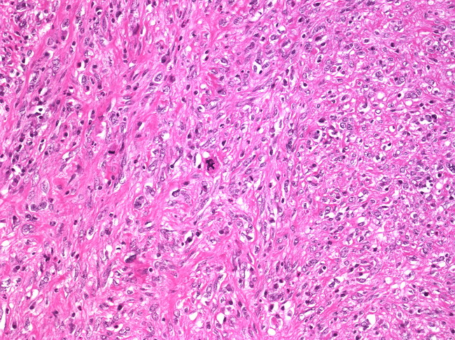
Malignant Fibrous Histiocytoma of Breast
slide 1 of 2


Comments:
The patient was a 60 y/o woman who presented with a 6.0 cm right breast mass. The tumor had a somewhat gritty cut surface. Twenty right axillary lymph nodes were negative for metastases. The tumor is composed of spindle cells arranged with a storiform growth pattern. There is marked nuclear pleomorphism and atypical mitotic figures are present. Chronic inflammatory infiltrate is present in the background.
slide 1 of 2