
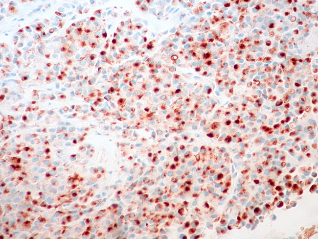
Aberrant E-Cadherin in Lobular CA

slide 34 of 55


Comments:
Most cases of in-situ and invasive lobular carcinoma (ILC) show lack of E-Cadherin immunoreactivity. However, aberrant staining is seen about 16% of cases and does not exclude the diagnosis of ILC if the H&E features are diagnostic. This image shows E-cadherin expression in the cytoplasm, possibly in the Golgi apparatus, and in the membrane bound intracytoplasmic vacuoles. The normal E-Cadherin staining (for e.g. in ductal carcinoma) is membranous. Image courtesy of: Dr. Ed Uthman, Houston, USA.
slide 34 of 55