
Pancreatoblastoma : Microscopic


Comments:
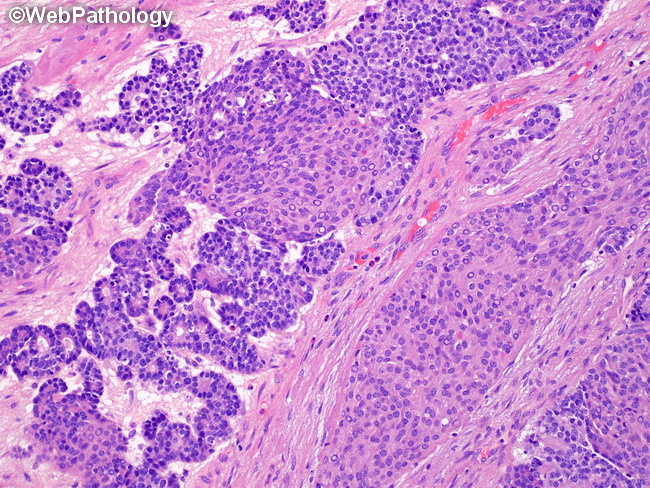
Microscopic Features: Pancreatoblastomas try to recapitulate early pancreatic development during embryogenesis, like many other blastomatous tumors of childhood. The histologic components show several lines of differentiation, including acinar, ductal, endocrine, as well as squamous. The tumor is composed tightly packed lobules separated by fibrous bands. Within the lobules, the cells are arranged in acinar structures, rosettes, solid sheets, or trabecular patterns. The most abundant component is sheets of polygonal cells with distinct cell borders, pale basophilic cytoplasm, central round or oval nucleus with a single nucleolus, and variable mitotic activity. Acinar structures are often seen at the periphery of the sheets. They enclose small lumens with eosinophilic secretions. The radially-arranged cells have uniform basal nuclei, punctate nucleoli, and eosinophilic granular cytoplasm. There is no cytologic atypia. Mitotic activity is slightly increased. This image also shows two large squamoid nests (more on squamoid component in the next slide).