
Liver Metastases : Gross Pathology


Comments:
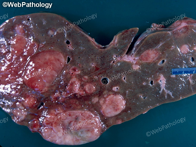
Gross Pathology: Liver metastases are usually multiple and form discrete expansive masses or ill-defined infiltrative areas that may greatly enlarge the liver. Larger lesion may undergo central necrosis and fibrosis producing umbilication. Metastatic deposits may be totally buried within the parenchyma and the external surface of the liver may be completely normal. Metastases are frequently gray-white with scattered areas of hemorrhage and necrosis. Individual nodules may be surrounded by areas of venous stasis. Metastases from highly vascular tumors such angiosarcoma, choriocarcinoma, and thyroid carcinoma are extensively hemorrhagic. Metastatic deposits from colo-rectal cancer may have a fibrous pseudocapsule and frequently undergo calcification if there is copious mucin production. Well-differentiated squamous cell carcinomas produce soft metastases due to necrosis and keratin production. Malignancies of breast, stomach, and pancreas sometimes produce miliary involvement with extremely small lesions scattered throughout the liver. It has been observed in autopsy studies that metastases are very rare in cirrhotic livers. Perhaps, the extensive scarring does not offer a hospitable, nutrient-rich background for the growth of extrahepatic malignancies. Grossly, many benign lesions can simulate metastases, including fibrous scars, healed granulomas, bile duct hamartoma, and focal nodular hyperplasia. Therefore, gross impression should not form the basis for the diagnosis of metastases by the surgeon intraoperatively or by the pathologist. The photograph shows liver metastases from a well-differentiated pancreatic neuroendocrine tumor.