
Metastatic Neuroendocrine Tumor


Comments:
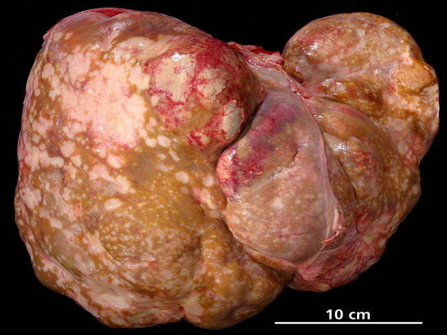
Primary neuroendocrine neoplasms (NENs) of the liver are extremely rare and much less common than liver metastases from NENs arising elsewhere in the gastrointestinal tract, pancreas, lung, or other sites. Therefore, before a NEN is accepted as a liver primary, the possibility of metastases must be excluded. The route of spread of NENs of gastrointestinal tract and pancreas to the liver is via portal circulation. The spectrum of metastatic neuroendocrine tumors in the liver is wide. It includes well-differentiated tumors (carcinoids) as well as small cell carcinomas. The well-differentiated tumors have a monomorphic appearance, minimal mitotic activity, and no necrosis on needle core biopsies. The smears are quite cellular with a monomorphic, often plasmacytoid appearance. Both smears as well as core biopsy samples show characteristic salt and pepper chromatin. High-grade tumors, such as small cell carcinomas from the lung, show nuclear molding, necrosis, increased mitotic activity, and finely stippled chromatin. Immunohistochemical panel consisting of synaptophysin, chromogranin, and CD56 is useful in confirming the diagnosis. Hepatic metastases from NENs are indolent and often highly symptomatic (Carcinoid syndrome). Complete resection often cannot be performed due to diffuse and multifocal involvement of the liver. Subtotal resection (debulking) is still recommended and can produce dramatic relief from symptoms of hormonal overproduction. The photograph shows extensive liver metastases from neuroendocrine carcinoma of pancreas. The patient developed hepatic encephalopathy and acute renal failure. Image copyright: pathorama.ch