
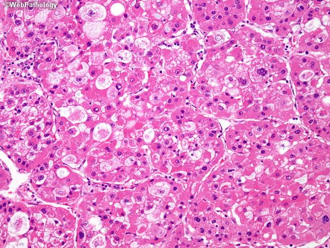
Hepatocellular Carcinoma - Pseudoglandular (Acinar) Pattern
Home
 Gastrointestinal
Liver
Liver Tumors & Tumor-like Lesions - II
Hepatocellular Carcinoma - Pseudoglandular (Acinar) Pattern
Gastrointestinal
Liver
Liver Tumors & Tumor-like Lesions - II
Hepatocellular Carcinoma - Pseudoglandular (Acinar) Pattern

slide 44 of 65


Comments:
The WHO describes several HCC histologic patterns, including trabecular (most common), acinar, solid, and scirrhous. Predominantly trabecular (sinusoidal) pattern HCC may also contain acinar (pseudoglandular) areas, as seen here. The tumor cells recapitulate native hepatocytes, with round to oval nuclei and abundant granular eosinophilic cytoplasm. The pseudoglandular spaces may occur secondary to central trabecular necrosis, whereupon they contain protein, cellular debris, or macrophages. Note the nuclear pleomorphism and prominent endothelial cell wrapping around tumor cell nests and trabeculae.
slide 44 of 65