
Hepatoblastoma - Teratoid Pattern
Home
 Gastrointestinal
Liver
Liver Tumors & Tumor-like Lesions - II
Hepatoblastoma - Teratoid Pattern
Gastrointestinal
Liver
Liver Tumors & Tumor-like Lesions - II
Hepatoblastoma - Teratoid Pattern

slide 18 of 65


Comments:
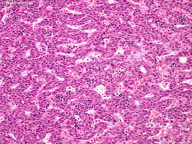
As in the previous case, this mixed epithelial-mesenchymal hepatoblastoma contains teratoid foci composed of tubulo-ductular structures of primitive epithelium with neuroectodermal features. The small dark cells scattered throughout the epithelium represent foci of extramedullary hematopoiesis, which is common in epithelial type hepatoblastomas. The patient was 1-day old and was found to have a large liver mass.
slide 18 of 65