
Liver Hemangioma : Imaging

slide 7 of 63


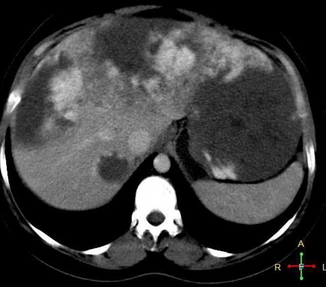
Comments:
Imaging: Being highly vascular lesions, liver hemangiomas show contrast enhancement on CT. During the arterial phase, there is peripheral, nodular, discontinuous enhancement. During the portal venous phase, there is progressive peripheral enhancement with centripetal fill-in which may be incomplete or irregular due to scarring (as shown here). Case courtesy of Dr Ahmed Abd Rabou, Radiopaedia.org. From the case rID: 22863
slide 7 of 63