
Dysplasia in Sessile Serrated Adenoma


Comments:
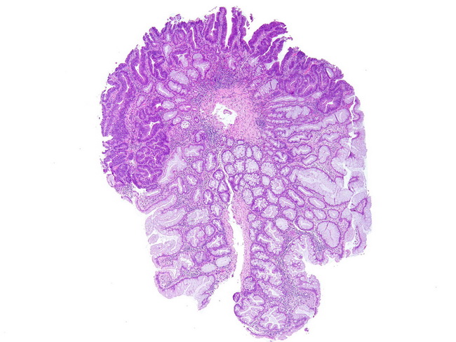
A small proportion of sessile serrated adenomas (SSA) show true cytologic dysplasia and contain areas resembling tubular adenoma (as seen on the top of this image). There is glandular overcrowding, nuclear enlargement and hyperchromasia, nuclear pseudostratification, and loss of goblet cell mucin. The dysplasia ranges from low-grade (most common) to high-grade and even invasive carcinoma (less common). The prevalence of dysplasia and carcinoma in SSAs ranges from 5% to 16%. The dysplastic foci usually show loss of MLH1 staining by immunohistochemistry and BRAF mutations. MLH1 is one of the genes involved in DNA mismatch repair. Image courtesy of: American Registry of Pathology Press (@ARP_Press); Used with permission.