
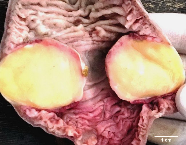
Lipoma


Comments:
This specimen is from a middle-aged female who presented with acute abdominal pain and nausea. CT abdomen-pelvis showed ileo-ileal intussusception measuring 4.6 cm x 4.4 cm in size and a hypodense lesion on the leading edge measuring 2.4 cm x 2.8 cm in size. Emergency laparotomy was undertaken and the involved segment of small bowel was resected. The specimen measured 14 cm in length and showed bulging and distension of about 4 cm diameter near one of the surgical margins. The bowel was opened longitudinally and showed a well-circumscribed sessile yellow tumor partially occluding the lumen. Microscopic examination confirmed it to be a lipoma. Intestinal lipoma presenting as intraluminal polyp is uncommon. Case courtesy of: Dr. Sanjay D. Deshmukh (Professor of Pathology) and Dr. Jayant M. Gadekar (Professor and Head of Surgery Dept.), Dr. Vithalrao Vikhe Patil Foundation's Medical College & Hospitals, Ahmednagar, India.