
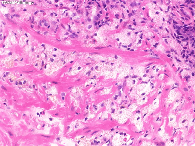
Prostatic Xanthoma

slide 48 of 58


Comments:
The lipid-laden histiocytes have vacuolated cytoplasm containing light brown pigment. The nuclei are small and there are no prominent nucleoli. Distinction from hypernephroid pattern of high-grade prostatic adenocarcinoma can usually be done without difficulty in most cases based on H&E features alone.
slide 48 of 58