Comments:
The following diagnostic categories are used in the context of Barrett esophagus (BE).
- Negative for Dysplasia
- Indefinite for Dysplasia
- Positive for Low-grade Dysplasia
- Positive for High-grade Dysplasia
- Intramucosal Carcinoma
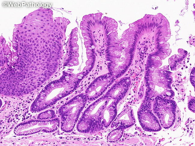
Negative for Dysplasia: This image shows BE, negative for dysplasia. There is
normal surface maturation from the basal glands to the top. The nuclei in the basal portion are slightly larger and more hyperchromatic than those at the surface. Overall, there is
no cytologic atypia. The nuclei have smooth contours. Nucleoli are not enlarged.
Nuclear polarity is
maintained throughout the glands. Some degree of nuclear stratification, a few mitoses in the basal glands, and a few dysmorphic goblet cells are acceptable. The
glandular architecture is
uniform. There is
abundant lamina propria separating the glands (i.e. no overcrowding). In the presence of ulceration, erosion, or acute inflammation, there may be reparative changes. The nuclei may become enlarged and hyperchromatic with prominent nucleoli, but should still maintain smooth contours. Surface maturation should be normal. There may be some loss of surface mucin.