
Toxoplasma Lymphadenitis
Home
 Hematopathology
Lymph Node (Non-Hematopoietic)
Protozoal Lymphadenitides
Toxoplasma Lymphadenitis
Hematopathology
Lymph Node (Non-Hematopoietic)
Protozoal Lymphadenitides
Toxoplasma Lymphadenitis

slide 5 of 7


Comments:
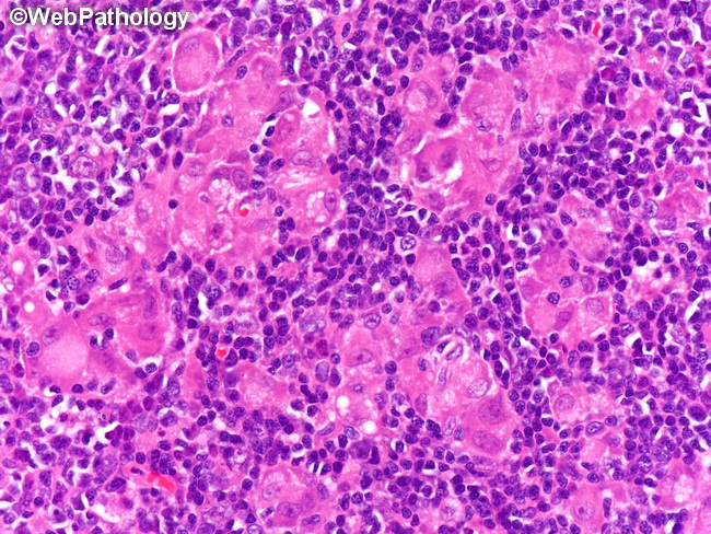
Toxoplasma Lymphadenitis: The epithelioid histiocytes are scattered singly and in small aggregates in the cortex and paracortex. They frequently encroach hyperplastic follicles, blurring their margins. The cells have abundant pale pink cytoplasm, oval nuclei with one or two punctate nucleoli, and no mitoses. There are no organized, well-formed granulomas or multi-nucleated giant cells. Necrosis or fibrosis are not seen.
slide 5 of 7