
Condyloma Acuminata : Microscopic
Home
 DermPath
Non-Neoplastic DermPath - II
Skin Infections - Viral
Condyloma Acuminata : Microscopic
DermPath
Non-Neoplastic DermPath - II
Skin Infections - Viral
Condyloma Acuminata : Microscopic

slide 31 of 56


Comments:
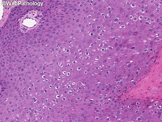
Microscopic Features: Condylomata are proliferative exophytic lesions of squamous epithelium with an orderly maturation. There is marked acanthosis, papillomatosis, hyperkeratosis, and parakeratosis. Superficial layers show vacuolated keratinocytes (koilocytes) and prominent coarse keratohyalin granules. Binucleated cells, dyskeratocytes, and wrinkled nuclei (resinoid appearance) may also be seen.
slide 31 of 56