
Cellular Neurothekeoma : Microscopic


Comments:
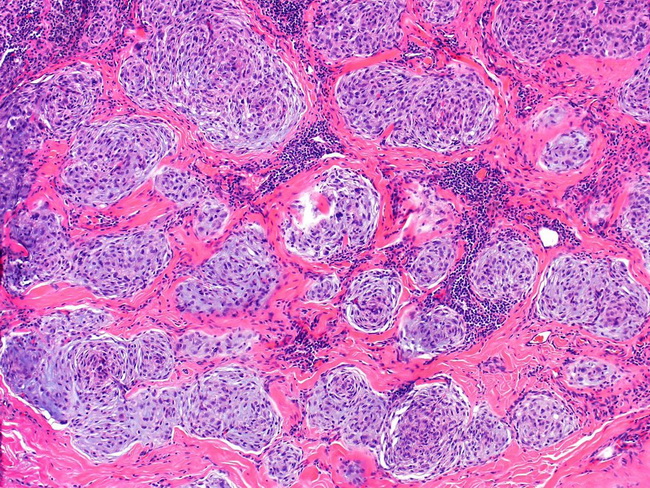
Microscopic Features: Neurothekeomas show multinodular, lobular, or plexiform growth patterns. They are composed of epithelioid or spindle cells arranged in round or oval nests of varying sizes separated by dense collagen bands. Individual nests frequently show whorling pattern. Some cases grow in solid sheets. The tumor cells have uniform ovoid nuclei with punctate nucleoli and lightly eosinophilic cytoplasm. Some cases can show moderate or even marked cytologic atypia. About 20% of cases show high mitotic activity and some may even have atypical mitoses. These atypical features are not a sign of malignancy and have not been associated with worse outcome. Osteoclast-like giant cells are present in about 40% of cases. The background stroma has myxoid foci, which, in some cases can be quite prominent and mimic other myxoid soft tissue tumors such as nerve sheath myxoma, myxofibrosarcoma. Image courtesy of: Rami Al-Rohil, MBBS, Department of Pathology, Duke University School of Medicine, Durham, North Carolina, USA; used with permission.