
Hyaline-Vascular Castleman Disease : Microscopic


Comments:
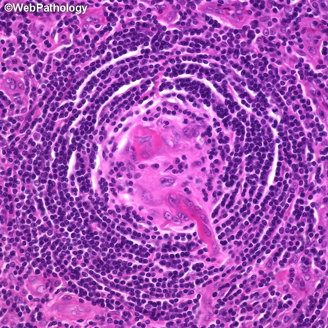
Microscopic Features of Hyaline-Vascular Castleman Disease (HVCD) (continued): The hyalinized cores of the germinal centers are composed mostly of CD21+ and CD35+ follicular dendritic cells (FDC), endothelial cells of proliferating vessels and scant residual follicle-center B-cells. The mantle zone is expanded and consists of several concentric layers of CD20+ B lymphocytes around the follicles, creating an onion-skin appearance. Blood vessels may penetrate the germinal center at right angles and give a "lollipop" appearance to the follicle. More than one germinal center may be present within a single mantle (twinning). Outside the follicles, the interfollicular region is greatly expanded by proliferation of high endothelial venules and a mixed cell population of plasma cells, eosinophils, plasmacytoid dendritic cells (CD123+), and TdT+ T-cells.