
Whipple Disease Lymphadenitis : Microscopic


Comments:
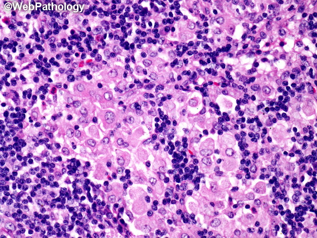
Microscopic Features of Whipple Disease: There is usually marked enlargement of mesenteric and periaortic lymph nodes in Whipple disease. The nodal architecture is distorted by numerous poorly-formed lipophagic granulomas and cystic spaces. Epithelioid and foreign body giant cells may be present, but there is no necrosis. There are aggregates of foamy histiocytes (as shown here) containing PAS-positive diastase-resistant material which is most likely residue of partially-digested bacilli. The foamy granular material is also gram-positive and silver (GMS stain) positive and acid-fast negative (an important distinguishing feature from MAI granulomas). Rare cases show monoclonal B-cell proliferation or even frank lymphomas. The duodenum and jejunum develop plaque-like lesions, blunted villi, and dilated lymphatics. Like lymph nodes, the intestinal lamina propria also contains aggregates of foamy macrophages filled with intracytoplasmic PAS-positive granules. In disseminated disease, such macrophages can also be seen in endocardium, pericardium, liver, spleen, synovium, mesentery, meninges, cerebral cortex, adrenals, and muscles.