
Kimura Disease : Differential Diagnosis


Comments:
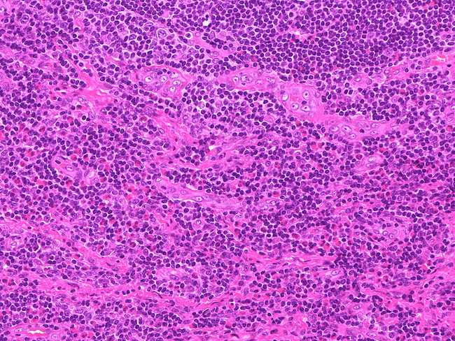
The differential diagnosis of Kimura disease includes: 1) other conditions with a prominent eosinophilic infiltrate such as angiolymphoid hyperplasia with eosinophilia (now renamed epithelioid hemangioma), Hodgkin lymphoma (mixed cellularity type), peripheral T-cell lymphomas, parasitic infections, drug reactions and histiocytosis X; and 2) conditions with follicular hyperplasia such as Castleman disease and dermatopathic lymphadenitis. Kimura Disease vs Angiolymphoid Hyperplasia with Eosinophilia (ALHE): Kimura disease and ALHE were previously considered to be related and/or represent different ends of spectrum of the same disease. Kimura disease was thought to be late stage of ALHE. However, studies have shown that they are distinct entities and ALHE has been renamed epithelioid hemangioma to reinforce this notion. ALHE is more common in Caucasian females. It is largely limited to the superficial dermis and is usually not associated with regional lymphadenopathy. The vascular channels are lined by plump epithelioid endothelial cells which may occlude the lumens. Kimura disease lacks epithelioid endothelial cells which are a diagnostic hallmark of ALHE. Lymph node involvement, peripheral blood eosinophilia, eosinophilic microabscesses, and elevated serum IgE are more common in the setting of Kimura disease. The image shows proliferation of post-capillary venules and eosinophilic infiltrate in the paracortical area of a lymph node affected by Kimura disease. Image courtesy of: @Patholwalker