
Lymphocyte Depleted Hodgkin Lymphoma
Home
 Hematopathology
Mature B-cell Neoplasms - Part I
Classic Hodgkin Lymphoma
Lymphocyte Depleted Hodgkin Lymphoma
Hematopathology
Mature B-cell Neoplasms - Part I
Classic Hodgkin Lymphoma
Lymphocyte Depleted Hodgkin Lymphoma

slide 72 of 84


Comments:
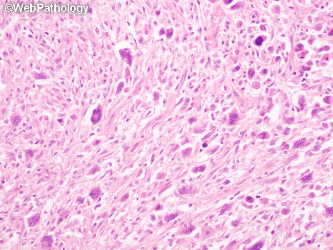
Higher magnification of the previous image showing lymphocyte-depleted classic Hodgkin lymphoma (cHL), diffuse fibrosis type. There are numerous atypical Reed-Sternberg (RS) cells in a background with sparse inflammatory infiltrate, only a few small lymphocytes, and heavy fibrosis. Some of the RS cells have a sarcomatous appearance.This variant of cHL may be difficult to distinguish from anaplastic large cell lymphoma on H&E alone. Cases with organized, concentric collagen bands around cellular nodules should be diagnosed as nodular sclerosis cHL.
slide 72 of 84